
Best Pleural Disease Treatment in Delhi
The pleura is the membrane that lines the thoracic (chest) cavity and covers the lungs. It is like a large sheet of tissue that wraps around the lungs’ outside and lines the chest cavity’s inside. There are several types of pleural diseases, including:
- Pleurisy – an infection of the pleural cavity
- Pleural effusion – the buildup of pleural fluid in the pleural cavity
- Pneumothorax – the presence of air or gas in the pleural cavity
- Hemothorax – the presence of blood in the pleural cavity
- Pleural tumours
What are the Symptoms of Pleural Diseases?
Symptoms of pleurisy may include:
- Shortness of breath
- A cough
- Fever and chills
- Rapid, shallow breathing
- Unexplained weight loss
- Sore throat that is followed by joint swelling and soreness
Symptoms of Pleural Effusion
Typically, pleural effusion causes no symptoms
Symptoms of pneumothorax include:
- Sudden sharp pain that worsens with deep breathing
- Shortness of breath
- Chest tightness
- Fatigue
- Fast heart rate
- Bluish skin color (called cyanosis)
Symptoms of hemothorax may include:
- Chest pain
- Shortness of breath
- Respiratory failure
- A rapid heart rate
- Anxiety
- Restlessness
Symptoms of pleural tumours may include:
- Shortness of breath
- Chest pain
- General discomfort
- Cough
- Unexplained weight loss
Causes of Pleural Disease
Causes of pleurisy include:
- Viral, bacterial, and fungal infections
- Lung cancer
- Other lung diseases, such as sarcoidosis, asbestosis, lymphangioleiomyomatosis, and mesothelioma
- Pulmonary embolism
- Familial Mediterranean fever
- Parasites
- Heart surgery
- Chest injury (trauma)
- Reaction to certain medications
Causes of pleural effusion include:
- Congestive heart failure
- Lung cancer
- Pneumonia
- Tuberculosis, asbestosis, sarcoidosis, and reactions to medications
- Bulla, which is a sizeable distended air space
- Lung diseases such as chronic obstructive pulmonary disease
- Tuberculosis
- Surgery
- Trauma
- Chest trauma
- Lung and pleural cancer
- Chest or heart surgery
Causes of hemothorax include:
For some pleural tumours, the cause is unknown. Known causes of pleural tumours may include cancer that has spread to the pleural space.
Treatment
After a careful evaluation, the medical team will recommend the appropriate treatment for each patient’s particular circumstances. Depending on the pleural condition and its cause, treatment may include:
- Bullectomy: Bullectomy is the surgical removal of a bulla, an air pocket in the lung that is greater than one centimetre in diameter (across). Bullae tend to occur as a result of lung tissue destruction and diseases such as cancer and emphysema. Their presence in the lung takes up space, causes pressure and blocks your breathing.
- Chemotherapy: Chemotherapy is a medication or combination of medications used to treat cancer. Chemotherapy can be given orally (as a pill) or injected intravenously (IV). When chemotherapy drugs enter the bloodstream, they destroy cancer cells. Chemotherapy is beneficial for cancers that have metastasized, or spread. Chemotherapy attacks all quickly-dividing cells, regardless of whether they are cancerous which can cause several side effects, including hair loss, mouth sores, loss of appetite, nausea and vomiting, diarrhoea, and low blood counts. Low blood counts can increase a patient’s risk of infection, bruising or bleeding, fatigue, and shortness of breath. The side effects of chemotherapy are generally temporary and often go away once treatment is completed. Chemotherapy regimens vary from patient to patient. They are generally repeated several times in cycles, with three to four weeks separating each cycle to allow damaged normal cells time to recover. After the first two or three chemotherapy sessions, patients may have a CT or PET scan to see if the drug(s) is effective. If the drug(s) is not working, it may be switched out for a new drug(s).
- Minimally Invasive Tumor Removal
- Video-Assisted Thoracoscopic Surgery (VATS) is a minimally invasive alternative to open-chest surgery that involves less pain and recovery time. After giving you a sedative, the physician will make tiny incisions in your chest and then insert a fibre-optic camera called a thoracoscope as well as surgical instruments. As the physician moves the thoracoscope around, images that provide important information are projected on a video monitor. VATS is not appropriate for all patients; you should have a thorough discussion with your provider before deciding. It is often not recommended in people who have had chest surgery in the past, because remaining scar tissue can make accessing the chest cavity more challenging and thus riskier.
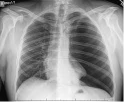
- Pleurodesis / Pleural Effusion: Pleurodesis is a therapy that a lung cancer doctor offers patients to remove excess fluid—called pleural effusion—from the space between the lungs and chest wall that line the lungs (pleura). This fluid prevents the lungs from fully expanding as you breathe, causing shortness of breath. Pleural effusion is usually diagnosed using a chest x-ray, and doctors may take a sample of the fluid to confirm its cause. There are a few ways to perform pleurodesis. One such way is video-assisted thoracoscopy, a new, less invasive method that we offer at BMC. Using a thoracoscope, a small, thin instrument with a light and lens, your surgeon will locate the area to be treated, drain your lung fluid, and then insert a talcum powder or antibiotic solution. This solution will circulate in the space between the pleura lining the chest wall and the lungs, causing some minor irritation and inflammation, which then causes the tissues to stick together, eliminating the space. Further fluid buildup is thereby prevented, allowing you to breathe easier. If the procedure is not successful, it may be repeated. Pleurodesis does not treat lung cancer, but it can be a beneficial tool in reducing symptoms.
- PleurX Catheter: The PleurX catheter is a thin, flexible tube placed in the pleural space to drain the fluid buildup associated with pleural effusion. Traditionally, treatment for chronic pleural effusion has required patients to remain in the hospital. The PleurX catheter allows you to manage your pleural effusion at home. The device consists of a catheter that is placed in the pleural space through a small incision. The catheter is connected to a vacuum bottle. When you open a valve at the end of the catheter, fluid drains into the vacuum bottle.
- Radiation Therapy: Radiation uses special equipment to deliver high-energy particles, such as x-rays, gamma rays, electron beams or protons, to kill or damage cancer cells. Radiation (also called radiotherapy, irradiation, or x-ray therapy) can be delivered internally through seed implantation or externally using linear accelerators (called external beam radiotherapy, or EBRT). Radiation may be used as a solitary treatment or with surgery and chemotherapy. The equipment used to deliver radiation therapy is called a linear accelerator. The linear accelerator has a moveable arm, enabling the radiation to be focused on the part of your body where the cancer is located. Developments in EBRT equipment have enabled physicians to offer conformal radiation. With conformal radiation, computer software uses imaging scans to map the cancer three-dimensionally. The radiation beams are then shaped to conform, or match, the shape of the tumour.
Radiation works by breaking a portion of a cancer cell’s DNA, which prevents it from dividing and growing. Radiation therapy can be systemic, meaning it moves throughout your bloodstream. Systemic therapies are usually given as an injection into a blood vessel or are taken as a pill. Systemic treatments expose your entire body to cancer-fighting medication. However, radiation therapy is typically given as a “local” treatment, meaning it affects only the part of the body that needs therapy. - Thoracentesis: The removal of pleural fluid with a needle or catheter that your thoracic surgeon inserts through your ribs in the back of your chest into your chest wall.
- Thoracostomy: During thoracostomy, the physician will inject a local anaesthetic into the chest wall where the fluid is located and place a plastic tube into the chest between two ribs. The physician will then connect the tube to a suction device, which will help to remove the fluid
- Tumour Ablation: Tumor ablation is an image-guided, minimally invasive treatment used to destroy cancer cells. A physician inserts a specially equipped needle (probe) into the tumour or tumours guided by computed tomography (CT) in tumour ablation. Once the probe is in place, energy is transmitted through it and into the tumour